Call Us Now1-800-414-2174


Medical Tip
Throughout our clinical career, Trevor and I have examined and treated numerous patients with low back pain (LBP) who were considering having back surgery when they did not actually need it. On many of those occasions, the basis for wanting back surgery was related to a so called “terrible finding” on a MRI. The “terrible findings” reported by patients included degenerative disc disease, spondylosis (degenerative vertebra), the dreaded “bulging disc”, spinal stenosis, facet arthropathy, etc. However, in too many cases we have found that the nature and/or severity of pain and disability was out of proportion to clinical findings, especially in litigation cases. Yet, the MRI was often relied on to be the “gold standard” to validate their complaints.
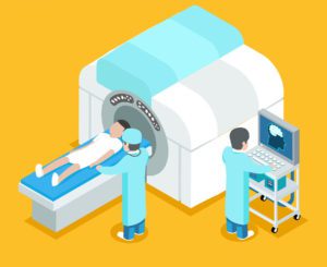
There is no doubt that magnetic resonance imaging (MRI) has been an important medical diagnostic instrument since its inception in 1977. However, it is well known that MRIs can reveal images of pathology that are not symptomatic. It is tempting in an era of “rushed medicine” to correlate an MRI finding to symptoms reported. However, this can be a costly mistake. Studies indicate that people walking the street every day without any appreciable back pain can have signs of pathology show up on MRI. In fact, in a study by Bolden, et al, it is opined that over 57% of working population would have “abnormal findings” if they undergo a lumbar MRI.1 Other studies on MRI findings of subjects who had no reports of symptoms have shown a high incidence of bulging discs, disc protrusions, and annual tears at one or more spinal levels.6,7,8
Although patients may request an MRI to satisfy their fears regarding the source of their LBP, those who receive an adequate explanation for their symptoms are less likely to want additional diagnostic tests. When MRIs are conducted, to reduce fear of MRI results, patients may benefit from an explanation that describes how pathology demonstrated on MRI may not be relevant to their symptoms. In fact, it is rare that a person age 50 or older does not present with abnormalities in the lumbar spine.
When it comes to low back pain (LBP) studies indicate that radiographs (x-rays) and MRI studies are generally over-utilized.2,3,4,6 Most LBP fits into the category of a degenerative condition that typically follows a mechanical pattern of pain and improves significantly with proper treatment within four weeks thereby not requiring imaging and particularly not advanced imaging such as an MRI.5
When red flags or examination findings suggest the presence of a serious underlying spinal condition such as fracture, cancer, infection, or cauda equina syndrome, the need for more extensive diagnostic evaluation including MRI is indicated even if the initial radiographic findings are negative. The sensitivity of MRI for cancer is high, ranging from 83 to 93% with a specificity of 90 to 97%. For spinal infection, MRI is 96% sensitive and 92% specific with an overall diagnostic accuracy of 94%.3
The sensitivity and specificity of MRI for herniated discs is higher than computed tomography (CT) but very close to computed tomography for spinal stenosis.3 The clinical examination for patients with sciatica should include straight leg raising and neurologic testing. A straight leg raise test is more sensitive than specific for disc herniation with reported sensitivity of 91% and specificity of 26%. 9,10,11
Whenever a healthcare provider is considering an MRI for a back injury or condition, it is essential that the need for the imaging be derived from a carefully conducted patient examination. Advanced specialty practice examination principles should guide appropriate clinical decisions regarding back imaging. These principles include planning and performing an examination that is consistent with impressions derived from the patient interview, utilizing appropriate tests and measures, adequately disrobing the patient, carefully palpating all injured structures, examining remote areas for possible associated injuries, and examining structures that may be referring symptoms into the area of concern.
Gross diagnostic confusion can result from referred pain leading to MRIs of unrelated structures. Pain that simulates sciatica in the posterior thigh, for example, a condition often related to nerve compression from a ruptured or torn disc, may be referred instead from other sources such as inflamed facet joints in the lower back (L5-S1 level), piriformis muscle strains in the buttock, sacroiliac joint dysfunction, or from other systems such as the cardiovascular, genitourinary, or gastrointestinal systems. Obtaining an MRI of an area of referred pain is an expensive high definition study of unrelated structures that may obscure the true diagnosis.
The healthcare expert should use cumulative knowledge, clinical experience, examination findings, and response to interventions such as physical therapy or manual therapy to determine the need for additional testing including MRI. Findings from the comprehensive examination help establish the nature, severity and irritability of the symptoms and provides the necessary correlation to MRI results. In conjunction with medical doctors specializing in spinal care, a McKenzie Certified Physical Therapist or a physical therapist certified in manual therapy are good choices for non-invasive treatments of typical spinal conditions. These therapists have excellent skills in differential diagnoses and work with medical physicians specializing in spinal care to help a patient recover from various low back disorders without the need for surgery.
In summary, a MRI is an excellent diagnostic tool. However, MRI findings should be skillfully correlated to symptoms by a detailed, comprehensive clinical examination. Care should be taken to avoid a rush to judgement based only on a MRI and perhaps a rudimentary physical examination. There are various causes of back pain, the cause of which may not be apparent on MRI and may be erroneously attributed to pathology that exists but is not contributing to the symptoms. Always seek second opinions before submitting to a back surgery. A physical therapist whose skills are focused on hands-on biomechanical examinations of the spine is a good choice to find out if the pain pattern correlates to the findings of a MRI. In addition, such a clinician can often provide a conservative treatment program that can result in full recovery without the need for surgery.
References:
You must be logged in to post a comment.
WorkSaver Employee Testing Systems 478 Corporate Dr. Houma, LA 70360
![]()
Thanks for the wonderful article